
Dental Diagnostics Gdańsk
- Dentist Gdańsk
- Service
- Dental Check-ups
Dental Diagnostics Gdańsk – Technology That Transforms Treatment
Did you know that 70% of treatment success depends on the quality of diagnostics? At Prodent Gdańsk, we don’t guess – we see. 3D CBCT tomography shows the exact bone structure before implantation, an intraoral scanner eliminates unpleasant impressions, and a Leica microscope reveals problems invisible to the naked eye. Thanks to the latest Planmeca Promax diagnostic equipment, we offer imaging in the highest resolution with radiation doses lower than during a plane flight. All examinations are performed digitally – results are immediate, without waiting. Precise diagnostics guarantee that your treatment will be optimally planned, safely performed, and yield lasting results.


Dental Diagnostics Gdańsk – Precision and Modernity at Prodent
This is a modern method of orthodontic treatment, based on 3D scanning, digital therapy planning, and precise selection of braces or aligners. Instead of traditional impressions, we use digital scanners that create an accurate image of your dentition in minutes. Based on this, the orthodontist can plan the entire course of treatment before it even begins – including the appearance of the final result.
Accurate diagnostics are the foundation of effective dental treatment. At Prodent Gdańsk, we have a full range of the latest generation diagnostic facilities: a Planmeca Promax 3D CBCT tomograph, digital radiovisiography, a Leica microscope with 40x magnification, 3Shape and iTero intraoral scanners, and Prof. Gerber’s facial arch for joint diagnostics. All examinations are performed digitally – images are available immediately, in the highest resolution, with minimal radiation dose. Precise 3D diagnostics allow us to plan every procedure with millimeter-level accuracy, minimizing risk and guaranteeing a predictable outcome. From simple periapical X-rays to advanced computed tomography – we provide comprehensive diagnostics before every treatment.
Why Diagnostics at Prodent?
- Planmeca Promax 3D – the most modern tomograph with a full range of X-ray examinations.
- Minimal Doses – digital technology, patient safety.
- Immediate Results – images available instantly, high HD resolution.
- Leica Microscope – diagnostics with 40x magnification.
- 3Shape and iTero Intraoral Scanners – digital workflow without impressions, patient comfort.
- Gerber Facial Arch – advanced joint diagnostics.
- 40 Years of Experience + ISO – highest standards.
- Two Locations – Garnizon and Madison, convenient access.
Planmeca Promax 3D – Top-Class Diagnostics
The central point of our diagnostic facilities is the Planmeca Promax 3D device – one of the most modern radiovisiography units in the world. It enables all types of digital X-ray images to be taken with minimal patient radiation exposure and maximum image resolution.
- Digital Radiovisiography (RVG) – periapical X-rays of individual teeth performed directly at the dental chair in each office. The image appears on the HD monitor immediately, allowing the doctor to continuously assess tooth anatomy, quality of root canal filling, tightness of prosthetic restorations, presence of carious lesions, or root fractures. Essential during root canal treatment, caries diagnostics, evaluation of prosthetic work, and implant procedures.
- Orthopantomogram (OPG) – a panoramic X-ray covering the entire oral cavity, mandible, maxilla, maxillary sinuses, and all teeth. A basic examination before orthodontic, surgical, implantological consultations, and comprehensive prosthetic treatment. It allows assessment of the condition of all teeth and implants, position of impacted teeth (wisdom teeth), general bone condition, presence of inflammation at root apices, and condition of the sinus mucosa. Based on the OPG, the doctor writes an initial treatment plan.
- Cone Beam Computed Tomography (CBCT) 3D – three-dimensional imaging of anatomical structures with precision unavailable to traditional 2D X-rays. The doctor can view images in various planes (axial, sagittal, coronal), assess inflammation, cysts, bone structure and density, the mucous membrane lining the sinuses, correctness of root canal treatment, position of roots relative to nerves and sinuses. Essential before implantation for precise planning of implant position and size, for treating difficult curved roots, assessing root fractures, planning surgical guides for navigated implantology (fully guided surgery).
- Lateral Cephalometric X-rays – a specialized lateral X-ray of the skull essential before orthodontic, prosthetic, and implantological treatment. It allows for the calculation of all cephalometric measurements, assessment of skeletal class, facial profile, and the relationship of soft tissues to bone.
- Temporomandibular Joint X-rays – diagnostics of joint problems, assessment of condyle position and space for the articular disc. Necessary during prosthetic rehabilitation, especially when increasing vertical dimension (raising bite height).
Leica Microscope – Diagnostics and Treatment with Magnification
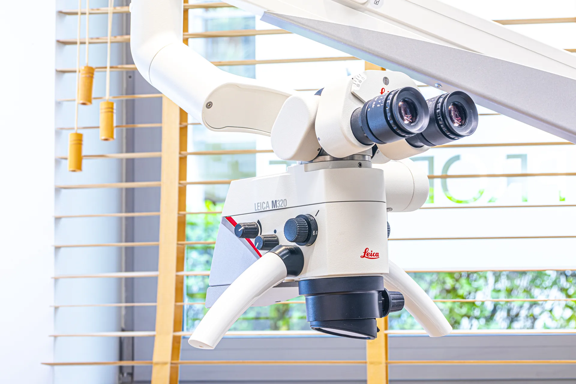
The Leica microscope with a vision track and 40x magnification is an indispensable tool in endodontics and prosthetics. An additional LED light source allows the doctor to see details invisible to the naked eye – additional root canals, microfractures, preparation margins. In prosthetics, it guarantees minimal tooth preparation and ideal sealing of veneers, onlays, and crowns according to the latest protocols. Working under magnification is the gold standard at Prodent.
3Shape and iTero Intraoral Scanners – Digital Precision at Your Service
The 3Shape TRIOS scanner is one of the most advanced devices on the market. It records a three-dimensional image of the dentition in real-time, eliminating the need for unpleasant silicone impressions. The scanner works with full color and tissue texture reproduction, allowing for precise planning of prosthetic, orthodontic, and implantological treatment. Data is sent directly to the dental laboratory in digital form, which shortens the turnaround time and minimizes the risk of errors.
The iTero scanner (Align Technology) has gained particular popularity due to its close integration with the Invisalign system. It not only enables precise reading of oral cavity anatomy but also simulates orthodontic treatment results – patients can see what their smile will look like after treatment before it even begins. iTero also features a function for monitoring the condition of the dentition over time – subsequent scans allow tracking changes and treatment progress.
Prof. Gerber’s Facial Arch – Joint Diagnostics
The dynamic facial arch is used for temporomandibular joint diagnostics and for making a distraction splint and wax-up in centric relation for prosthetic rehabilitation. We perform arch examinations for joint problems (clicking, tinnitus, disc displacement, tooth wear), before prosthetic work (veneers, crowns) for diagnosing occlusal disease, during orthodontic treatment when necessary, and before implantation.
Safety and Radiation Doses
Modern digital devices use radiation doses many times lower than traditional analog equipment. An orthopantomogram is approximately 0.02 mSv (equivalent to 2-3 days of natural background radiation), CBCT tomography is 0.05-0.08 mSv (about a week of natural background), a single RVG X-ray is only 0.001 mSv. For comparison: a flight from Warsaw to New York exposes one to 0.1 mSv. X-ray diagnostics at Prodent are completely safe.
Digital Archiving and Accessibility
All images taken at Prodent are digitally archived in a computer system and available immediately after being taken. Patients receive images in a convenient way – on a USB drive, by email, or via the cloud. We can also send them to other specialists (orthodontists, maxillofacial surgeons) for consultation. Digital documentation is durable, indestructible, and always accessible.
FAQ – Dental Diagnostics Gdańsk
1. Are X-rays safe?
2. What is the difference between 3D CBCT tomography and a traditional pantomogram?
3. When is 3D tomography needed?
4. Is tomography painful and how long does it take?
5. What is an intraoral scanner and what are its advantages?
The 3Shape TRIOS scanner is one of the most advanced devices on the market. It records a three-dimensional image of the dentition in real-time, eliminating the need for unpleasant silicone impressions. The scanner works with full color and tissue texture reproduction, allowing for precise planning of prosthetic, orthodontic, and implantological treatment. Data is sent directly to the dental laboratory in digital form, which shortens the turnaround time and minimizes the risk of errors.
The iTero scanner (Align Technology) has gained particular popularity due to its close integration with the Invisalign system. It not only enables precise reading of oral cavity anatomy but also simulates orthodontic treatment results – patients can see what their smile will look like after treatment before it even begins. iTero also features a function for monitoring the condition of the dentition over time – subsequent scans allow tracking changes and treatment progress.
6. Why does every Prodent office have RVG?
7. How to prepare for X-ray examinations?
8. Can I get my images on a storage device?
9. What is a facial arch and when is it needed?
10. How often can X-rays be performed?
OUR OFFER
Do you have dental problems? Contact us today!
Prodent Garnizon
+48 519 077 119
Opening Hours:
Prodent Madison
+48 519 077 118